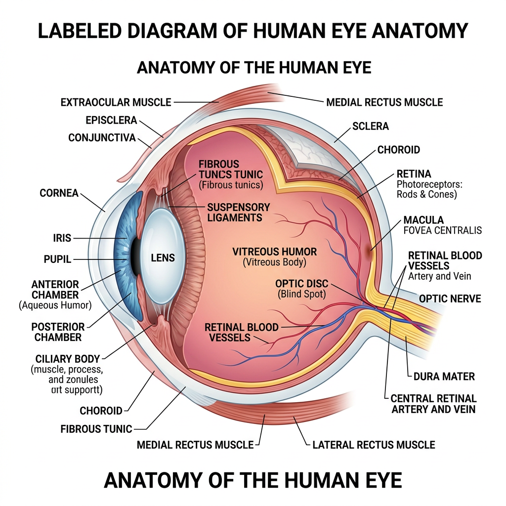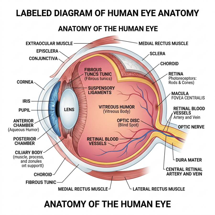
A labeled cross-sectional diagram showing the anatomy of the human eye.
The image is a detailed, labeled cross-sectional illustration of the human eye’s anatomy. It highlights key structures including the cornea, iris, pupil, lens, anterior and posterior chambers, ciliary body, fibrous tunic, choroid, retina, macula, optic disc, and optic nerve. The diagram also shows blood vessels such as retinal blood vessels and the central retinal artery and vein, along with muscles like the medial and lateral rectus muscles. The illustration uses clear labeling and color differentiation to visually separate parts, providing a comprehensive overview of the eye’s internal and external components.